|
|
|
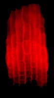
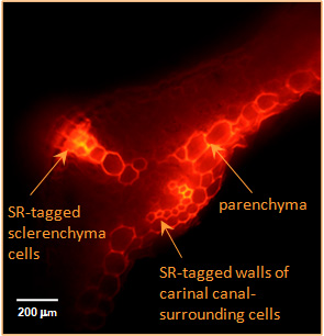
In-situ visualisation of transglycosylase action by the use of fluorescently labelled acceptor susbtratesTransglycanase action detected in the cell walls of living plant organs. Both the enzyme (XTH or HTG) and the donor substrate (xyloglucan and/or MLG) are endogenous to the organs. A small, fluorescently labelled acceptor substrate (xyloglucan-oligosaccharide–sulphorhodamine) was supplied exogenously. Fluorescence indicates the location of transglycanase action, creating polysaccharide–sulphorhodamine conjugates. Left: XET action in the elongation zone of an Arabidopsis root. Root tip (non-fluorescent) is at the bottom. (Photo: K Vissenberg 2003) Right: XET and MXE action in Equisetum fluviatile tissue. (Photo: L Franková 2011) |